Arm Muscles Diagram Anterior - Ct fletcher strongest man, healthy food to lose weight ... : Shoulder muscles chest muscles thigh muscles arm muscle anatomy muscle diagram bicep muscle yoga anatomy medical anatomy there are anterior muscles diagrams and posterior muscles diagrams.. Produce wrist and/or finger flexion. These are of course, anterior assuming the arm is in the anatomical position. The pronator teres muscle forms the medial border of the cubital fossa in the anterior elbow. Coracobrachialis is the most medial muscle in the anterior compartment of the arm. The deltoid consists three sets of fibers:
From anterior distal humerus d. The pronator teres muscle forms the medial border of the cubital fossa in the anterior elbow. The muscles of the upper arm are split into anterior and posterior compartments. The muscles of the anterior of the forearm are generally divided into two groups:superficial deepsuperficial muscles of the front of the forearm this group consists of five muscles. Flat muscle enabling various arm movements, such as drawing it near the median axis of the body and rotating it.
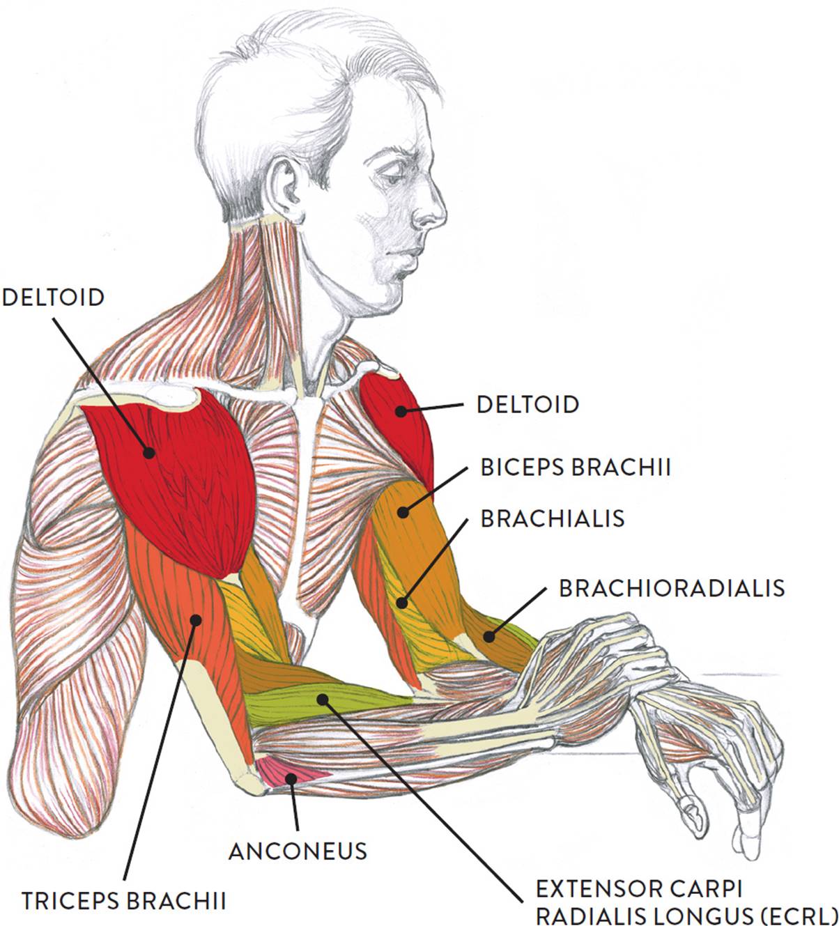
Muscles of Upper Extremity (Anterior Superficial view) from anatomyuniverse.com Adbucts scapula and rotates it downward. This layer contains only one muscle, the flexor digitorum. Usually as one muscle contracts (or shortens), the opposing muscle (known as the antagonist) elongates and vice versa. The muscles of the arm anatomical chart does an exemplary job of examining the individual muscles that make up this area of the human body, and included in the dozen or more beautiful illustrations are views of the dorsal area, thorax, triceps, biceps, brachialis, serratus anterior, promator and. Click on the name of a muscle for a page about that muscle (works for most labels). Produce wrist and/or finger flexion. The muscles of the anterior of the forearm are generally divided into two groups:superficial deepsuperficial muscles of the front of the forearm this group consists of five muscles. In this image, you will find hand and forearm muscle anatomy, humerus, extensor carpi radialis longus muscle, anconeous muscle, lateral antebrachial cutaneous nerve, ulna, radial nerve, flexor carpi ulnaris, median nerve, brachioradialis muscle, medial antebrachial cutaneous nerve in it.
Overview of anatomy and joint actions, with video, worksheets and common exercise analysis.
In this image, you will find hand and forearm muscle anatomy, humerus, extensor carpi radialis longus muscle, anconeous muscle, lateral antebrachial cutaneous nerve, ulna, radial nerve, flexor carpi ulnaris, median nerve, brachioradialis muscle, medial antebrachial cutaneous nerve in it. The muscles of the arm anatomical chart does an exemplary job of examining the individual muscles that make up this area of the human body, and included in the dozen or more beautiful illustrations are views of the dorsal area, thorax, triceps, biceps, brachialis, serratus anterior, promator and. Stabilizes the head of the humerus in glenoid cavity; Coracobrachialis brachialis biceps brachii coracobrachialis: Learn vocabulary, terms and more with flashcards, games and other study tools. Coronoid process and ulnar tuberosity the rough anterior. Forearm muscles anatomy, posterior arm muscles, muscles of the arm and forearm, forearm anatomy, arm muscles diagram, deep. Draw labelled diagram showing branches of profunda brachi artery. Anterior view, superficial muscles of the forearm. The anterior compartment contains three muscles; Get in touch with us today! Flat muscle enabling various arm movements, such as drawing it near the median axis of the body and rotating it. Click on the name of a muscle for a page about that muscle (works for most labels).
The muscles labelled in the anterior muscles diagram shown above are listed in bold in the following table The biceps brachii, the brachialis and the coracobrachialis. Ridge muscles of the arm. Anterior view, superficial muscles of the forearm. Usually as one muscle contracts (or shortens), the opposing muscle (known as the antagonist) elongates and vice versa.
Muscles of the Arm and Hand - Classic Human Anatomy in ... from doctorlib.info Draw labelled diagram showing branches of profunda brachi artery. The serratus anterior is a muscle that originates on the surface of the 1st to 8th ribs at the side of the chest and inserts along the entire anterior length of the medial border of the scapula. In general, these are the flexors of the wrist and fingers and pronate the forearm. The upper arm is located between the shoulder joint and elbow joint. Although the majority of the muscle mass is located anteriorly to the humerus, it has no attachment. The anterior compartment of the arm is also known as the flexor compartment of the arm as its main action is that of flexion. The biceps brachii, the brachialis and the coracobrachialis. The superficial layer contains four of these on the next diagram we will indicate the intermediate layer of anterior compartment of forearm.
It is a functionally important muscle that contains two heads.
These muscles are all innervated by the musculocutaneous. Learn the muscles of the arm with free quizzes, diagrams and worksheets. Flexion of the forearm is achieved by a group of three additionally, the biceps brachii operates as a supinator of the forearm by rotating the radius and moving the palm of the hand anteriorly. Muscles of anterior (flexor) compartment of arm, their origin, insertion, action/s and nerve supply are as follows superior ulnar collateral branch of brachial artery. Muscles of the arm anatomy, labeled diagram. The muscles of the upper arm are split into anterior and posterior compartments. The arm muscles are located between the shoulder and elbow joint. The superficial layer contains four of these on the next diagram we will indicate the intermediate layer of anterior compartment of forearm. Thick triangular muscle drawing the arm away from the median axis of the body and directing it toward the front and back until it is horizontal. Anterior view, superficial muscles of the forearm. Draw labelled diagram showing branches of profunda brachi artery. Tutorials and quizzes on muscles that act on the arm/humerus (arm muscles: Flat muscle enabling various arm movements, such as drawing it near the median axis of the body and rotating it.
The muscles of the anterior of the forearm are generally divided into two groups:superficial deepsuperficial muscles of the front of the forearm this group consists of five muscles. These are the pronator teres, flexor carpi radialis, palmaris longus, and flexor carpi ulnaris. Have a product modelling and rendering project?. Produce wrist and/or finger flexion. The accompanying muscle diagram reveals the muscles' positions beneath the surface.
My Blog: Muscles of the upper limb from lh4.googleusercontent.com You can see it running just underneath the biceps and it inserts onto the humerus. Name the muscle of extensor compartment of arm and its. These are of course, anterior assuming the arm is in the anatomical position. The anterior compartment is the flexor compartment because these we've just got a diagram of it here. The arm muscles comprise five muscles, which mainly act to flex and extend the forearm. This is the coracoid process. Start studying arm anterior muscles. Have a product modelling and rendering project?.
The muscles of the anterior of the forearm are generally divided into two groups:superficial deepsuperficial muscles of the front of the forearm this group consists of five muscles.
Arm anterior 3d illustration project. The deltoid consists three sets of fibers: The muscular system is made up of specialized cells called muscle fibers. Name the muscle of extensor compartment of arm and its. Coracobrachialis is the most medial muscle in the anterior compartment of the arm. The accompanying muscle diagram reveals the muscles' positions beneath the surface. In this image, you will find hand and forearm muscle anatomy, humerus, extensor carpi radialis longus muscle, anconeous muscle, lateral antebrachial cutaneous nerve, ulna, radial nerve, flexor carpi ulnaris, median nerve, brachioradialis muscle, medial antebrachial cutaneous nerve in it. Multiple muscles on the front of your arm shorten (biceps, brachialis, etc.) to allow for this to. Four main muscles of the anterior region of the lower arm begin on the medial epicondyle of the humerus. Usually as one muscle contracts (or shortens), the opposing muscle (known as the antagonist) elongates and vice versa. Arm anatomy diagram for artists. Introduction to functional anatomy of the arm muscles: Ridge muscles of the arm.
Biceps brachii, brachialis, triceps, brachioradialis and coracobrachialis arm muscles diagram. Supination of the forearm = lateral rotation of the radius such that the palm of the hand faces anteriorly.


Post a Comment
0 Comments